 Joe White is a fifth year student at The University of Sheffield. "I wanted to write this piece to share some rare experiences I had during my "non-orthodox" volunteering experience."
Joe shares tales from his volunteering period, shadowing veterinary maxillofacial surgeon - Dr Gerhard Steenkamp - in Pretoria, South Africa.
"Well, you couldn't have picked a better two weeks to visit me" was the welcome I received from veterinary maxillofacial surgeon and dentist Dr Gerhard Steenkamp as he flicked through the clinic diary on my first day at the University of Pretoria's Veterinary Campus. Dr Steenkamp runs a private referral clinic within the regional veterinary hospital, whilst also fulfilling his duties as a veterinary surgeon and lecturer for the university.
Veterinary Dentistry is a small specialty and is quoted on the BVDA website as "...an important, yet still underdeveloped area of veterinary practice." The main workload of every-day veterinary dentistry is management of periodontal disease and its sequelae with some endodontics, and even orthodontics, thrown in on the side. This appears to be mainly due to what some would consider neglect of pet oral hygiene, especially when "toy breed" dogs such as the Pug are more susceptible to periodontal disease and have grown in popularity in recent years.
The Elephant
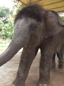
My second day involved an early start to be collected in Dr Steenkamp's pick-up truck, full of the most intimidating oral surgery set you have ever witnessed. We were on our way to a private reserve to see a 2-year long, ongoing case of a 30 year old bull elephant with a fractured left tusk.

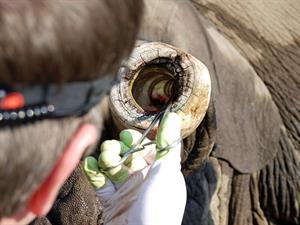
The keeper was complaining of chronic pus drainage from the tusk since the last appointment and had called Dr Steenkamp to come back to take a look. After anaesthetic induction with the most potent opioid known to man, Ten fully grown men had to guide the bull down onto the correct side for the procedure and also into a position where his internal organs (and delicate ears) wouldn't be crushed under his own weight. Dr Steenkamp started by accessing the infected pulp chamber through the necrotic debris and reactionary ivory that had formed, with metre-long Coupland's Elevators, a mallet, and a Black and Decker circular drill. Once all the infected ivory was removed and the access cavity was cleaned out with a garden hose, the hope was that a healthy, bleeding pulp would prevent itself. This would indicate that the pulp had indeed mounted an appropriate response to the infection and the tusk wouldn't require extraction - Dr Steenkamp's most dreaded procedure as you can imagine.
Once Dr Steenkamp was happy that the infected ivory had been removed, as all around looked on in amazement, the tusk orifice was packed with that infinitesimally cheaper brother of Mineral Trioxide Aggregate - Portland Cement straight out of the bag! This novel pulp cap was followed by a coronal seal consisting of a special designed nylon plug tapped into the "tusk canal". The nylon has a similar hardness as the ivory so with use, abrades down to ivory level to form a smooth margin. All that was left was for Dr Steenkamp to take me back to my accommodation on the beautiful Onderstepoort campus via his favourite "Biltong Joint" - a local form of dried seasoned meat; and one for everyone's bucket list!
Measuring outcomes
Measuring outcomes in this procedure, as with any veterinary procedure, is incredibly difficult. Think of how often your plans are changed by your patient complaining or mentioning something or shifting about as you blow the 3-in-1 between their pre-molars? Vets are going in blind and some very interesting techniques are employed to diagnose and measure success. When this elephant had originally presented, the exposed tusk was diagonally fractured and hence unrestorable and had to be amputated down to gingival/skin level. When this was done, it allowed the owner and Dr Steenkamp to measure healing simply through comparative growth as tusks are elodonts (constantly growing) teeth. If the fractured tuck was growing at the same rate as the contralateral, we can assume the apex is healthy and the tusk won't require extraction. Elodont teeth are found in many animals such as squirrels, rabbits, and elephants. They may be incisors, canines, molars, but they are all unified in that they have an open apex - usually deep in the skull - from which they grow as they are used. Interestingly, the reason squirrels/beavers have chisel shaped elodont incisors is because enamel is only formed on the labial surface and therefore, the dentine behind it abrades away faster to form a chisel shape, better suited for use! Dr Steenkamp was also investigating the occurrence of Elodontomas in Tree Squirrels. These are exactly as they sound, benign growths of aberrant dental tissue around the apex which in obligate nasal breathers such as squirrels, occludes the nasal cavity and eventually prove deadly.
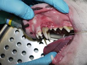
A couple of days later and we were in theatre, trying to stabilise a bilaterally dislocated TMJ in a seven year old cat. Unfortunately, the trauma had torn the TMJ capsule and surrounding ligaments so Dr Steenkamp had to devise a supportive nylon suture sling connecting the zygomatic arch and the condylar neck. The cat was then taken to his clinic for root canal treatment on all four fractured canines, and then placement of endodontic posts as the most predictable method of inter maxillary fixation in felines is bonding of the canines with composite resin.
My Last Day
My last day was marginally less scary then the preceding - standing alongside five other men, all holding onto a fire hose, looped around the neck of a poorly-sedated hippo, whilst Dr Steenkamp attempted to amputate a malocclused lower right canine with a power saw. We'd been called to a private clinic to see a rescued two year old wild leopard.

Dr Steenkamp was telling me that local practice by farmers involves large trap cages which can be left for days with leopards and other wildlife trapped inside. These wildcats often fracture numerous teeth and cause soft tissue trauma by trying to escape through the wire. Radiographs showed open apices on all four fractured canines, rendering Dr Steenkamp's methods of root filling, useless. The only option was to extract allfour, along with both upper third incisors - also fractured. The extraction of canines in cats and dogs is nothing like any of us can imagine. For starters, the roots are incredibly long and curved, and the pulp canals are so wide that forceps just crunch the tooth. Every single one has to be a surgical, not just because of the necessity of finding an application point on the tooth, but because oro-nasal communications occur in the maxilla with almost every canine extraction; as I was made aware when we couldn't stop a Staffordshire terrier projectile-sneezing blood clots all over the surgery.
One experience that was striking more than any other was the case of a two year old labrador with a melanoma infiltrating most of her left maxilla that had become so huge, she had begun to chew it. Many canine species have pigmented mucosa and this is not only due to more melanin pigment, but also more melanocytes. As one of the earliest cases I saw, it wasn't easy to take in. After reviewing a CT scan which would make any human surgeon's heart sink, surgery was scheduled for a few days later. You could see how much the dog meant to her owner and the lengths the client was willing to go to save the dog were truly heartwarming. The dog had most maxilla removed from the fourth premolar, caudally - that is posteriorly, in human terms; in addition to her entire coronoid process, which is incredibly large in dogs. Her eye had to be supported with a nylon sling from the remaining supra-orbital rim to the zygomatic buttress/infraorbital rim. The end result was amazing given what the dog had been through: she had one day of ketamine and walked out with her owner, eating and drinking normally, three days later.
I hope these tales have been interesting and if anyone is considering a voluntary placement out of the ordinary, then Dr Steenkamp welcomes veterinary and dentistry students alike. If anyone is interested, please contact me via email and I will put you in touch with him. For an immensely rewarding, relatively cheap (I had the pleasure of a superb T-bone steak and beer for the equivalent of £2.80 close to the hospital) an unforgettable experience, consider veterinary dentistry and I assure you, you won't be disappointed.
Joe White
[email protected]